Bi monthly exam Jan 16


 This is my submission for the Bimonthly internal assessment for the month of January ."

Most of the information here have been collected from different reference sites, the links to which have been mentioned. The points copy pasted have been put in quotes. 
The questions to the cases being discussed can be viewed from the link below ๐Ÿ‘‡


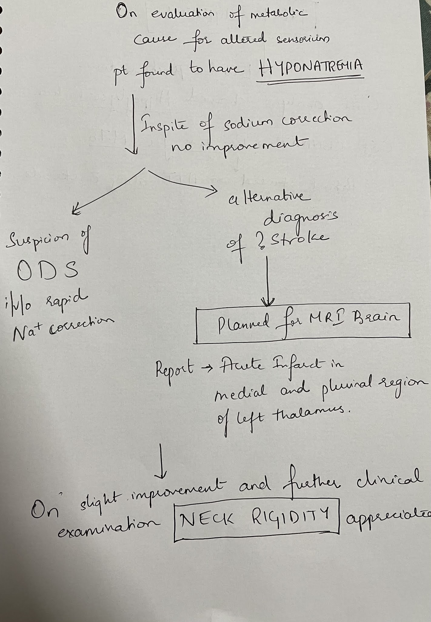
26 year old woman with complaints of altered sensorium somce 1 day,headache since 8 days,fever and vomitings since 4 days


a). What is the problem representation of this patient and what is the anatomical localization for her current problem based on the clinical findings?

Ans- The problem list in this patient can be illustrated as following
1) Altered sensorium on day of presentation to casualty
2) Motor weakness of all 4 limbs
3) Headache and neck pain from 8days
4) H/o joint pains involving small and large joints since 10yrs.

By analysing the sequence of symptoms she developed the anatomical localisation for her problems would point towards an immune mediated reaction involving central nervous system( Brain)and a separate entity for her skeletal complaints.


b) What is the etiology of the current problem and how would you as a member of the treating team arrive at a diagnosis? Please chart out the sequence of events timeline between the manifestations of each of her problems and current outcomes. 

Patient was diagnosed with 
Tubercular meningitis
Hyponatremia which can be attributed to SIADH secondary to meningitis( IL 1,TNF alpha,and IL 6 mediated release of ADH from post pitutary)


c) What is the efficacy of each of the drugs listed in her prior treatment plan that she was following since last two years before she stopped it two weeks back? 

HCQ use in SLE

It has been implicated that hcq causes reduction of serum levels of immunomodulators which are the culprits for clinical manifestations of sle

‘’Two-month treatment with HCQ resulted in significant decrease in SLEDAI-2K (p < 0.001), anti-dsDNA (p < 0.001), IL-1ฮฒ (p = 0.003), IL-6 (p < 0.001) and TNF-ฮฑ (p < 0.001) and a significant increase in CH50 levels (p = 0.012). ‘’


What is the efficacy of using primary bisphosphonate prophylaxis for patients started on corticosteroids?

Pooled analysis for incident nonvertebral fractures included nine trials with 1245 participants with low‐certainty evidence (downgraded for imprecision and serious risk of bias as a patient‐reported outcome). In this analysis 30/546 (or 55 per 1000) people experienced new nonvertebral fracture in the control group compared with 29/699 (or 42 per 1000; range 25 to 69) in the bisphosphonate group
A meta analysis showing there is no much difference in the outcomes as far as non vertebral fractures are concerned!


d) Please share any  reports around similar patients with SLE and TB meningitis?

Here's a case report of a 31 year old woman diagnosed with  SLE and subsequently developed SLE myocarditis and Lupus Nephritis who also developed TB meningitis


Another case report of a 51 year old woman with SLE who later developed TB meningitis

e) What is the sensitivity and specificity of ANA in the diagnosis of SLE? 

To determine the sensitivity and specificity of ANA and anti-dsDNA in SLE patients, using sera from Healthy controls and Multiple Medical problem patients.
The prevalence of ANA at a titer of ≥1:80 and ≥ 1:160 was 8% and 4%, respectively, in HC; and it was 12% and 6% respectively, in MMP patients. The prevalence of anti-dsDNA was 0% in HC and 3% in MMP patients. When using HC sera for the diagnosis of SLE, the sensitivity of ANA at a titer of ≥ 1:80 and ≥ 1:160 was 98% and 90%, respectively, with specificity of 92% and 96%, respectively. The specificity decreased to 88% and 94%, respectively, when using sera from MMP patients. The specificity of anti-dsDNA was 100% and 97%, when using sera from HC and MMP patients, respectively.
ANA and anti-dsDNA gave high sensitivity and high specificity in patients with SLE, even when using MMP patient's sera as controls.


2) Please go through the two thesis presentations below and answer the questions below by also discussing them with the presenters:


1)What was the research question in the above thesis presentation? 

To study the association of serum magnesium levels of type 2 diabetes mellitus


2) What was the researcher's hypothesis?

Normal magnesium levels are 1.6 to 2.4
Where as in diabetics substantial amount of population have a low magnesium levels in the blood, also in non diabetic populations, a low magnesium level is a danger sign that the person might develop diabetes mellitus

Magnesium deficiency is associated with hypocalcemia, hypokalemia and arrhythmias and hence correlates with a higher mortality /morbidity

 If There is reduced serum magnesium they have a negative impact on glucose homeostasis and insulin sensitivity,leading to early  development of complications such as nephropathy,neuropathy and retinopathy and thus have worst outcomes compared to people who are diabetic and have normal magnesium levels.
The diabetic complications progress faster in these populations


3) What is the current available evidence for magnesium deficiency leading to poorer outcomes in patients with diabetes? 

“Hypomagnesemia occurs at an incidence of 13.5 to 47.7% among patients with type 2 diabetes. Poor dietary intake, autonomic dysfunction, altered insulin metabolism, glomerular hyperfiltration, osmotic diuresis, recurrent metabolic acidosis, hypophosphatemia, and hypokalemia may be contributory. Hypomagnesemia has been linked to poor glycemic control, coronary artery diseases, hypertension, diabetic retinopathy, nephropathy, neuropathy, and foot ulcerations. The increased incidence of hypomagnesemia among patients with type 2 diabetes presumably is multifactorial. Because current data suggest adverse outcomes in association with hypomagnesemia, it is prudent to monitor magnesium routinely in this patient population and treat the condition whenever possible.”

 In addition to hyperosmolar coma and ketoacidosis, patients with type 2 diabetes may have cardiovascular disease, nephropathy, retinopathy, and polyneuropathy. With its associated complications, diabetes was reported to be the sixth leading cause of death. numerous studies have reported an inverse relationship between glycemic control and serum Mg levels. In patients with type 2 diabetes, oral Mg supplementation during a 16-wk period was suggested to improve insulin sensitivity and metabolic control

 There is a correlation between glycemic control and serum Mg levels or improvement of diabetic control with Mg replacement


Cardiovascular.

In a study that involved 19 normotensive individuals without diabetes, 17 hypertensive individuals without diabetes, and 6 hypertensive individuals with diabetes, Resnick et al. documented the lowest mean intracellular Mg concentration among the last group. Similarly, based on data from the Atherosclerosis Risk in Communities (ARIC) Study, a multicenter, prospective cohort study that lasted 4 to 7 yr and involved 13,922 middle-aged adults who were free of coronary heart disease at baseline, an inverse association between serum Mg and the risk for coronary heart disease was observed among men with diabetes 


Diabetic Retinopathy.

The link between hypomagnesemia and diabetic retinopathy was reported in two cross-sectional studies that involved both “insulin-dependent” patients and patients with type 2 diabetes. Not only did patients with diabetes have lower serum Mg levels compared with their counterparts without diabetes, but also the serum Mg levels among the cohort with diabetes had an inverse correlation with the degree of retinopathy .


Foot Ulcerations.

Given the link between hypomagnesemia and risk factors for the development of diabetic foot ulcers (e.g., polyneuropathy, platelet dysfunction), Rodriguez-Moran and Guerrero-Romero (48) suggested that hypomagnesemia may be associated with an increased risk of diabetic foot ulcers. Indeed, they observed a higher incidence of hypomagnesemia among their patients with diabetic foot ulcers compared with those without the condition (93.9% of the 33 patients with diabetic foot ulcers compared with 73.1% of the 66 patients without diabetic foot ulcers; P = 0.02).

Nephropathy.

In a comparative study that involved 30 patients who had type 2 diabetes without microalbuminuria, 30 with microalbuminuria, and 30 with overt proteinuria, Corsonello et al. (49) observed a significant decrease in serum ionized Mg in both the microalbuminuria and overt proteinuria groups compared with the nonmicroalbuminuric group. Accordingly, in a recent retrospective study, an association between low serum Mg levels and a significantly faster rate of renal function deterioration in patients with type 2 diabetes was reported (7).

Others.

Finally, there also are data to suggest the association between hypomagnesemia and other diabetic complications, including dyslipidemia and neurologic abnormalities.Because hypomagnesemia has been linked to various micro- and macrovascular complications, a better understanding of Mg metabolism and efforts to minimize hypomagnesemia in the routine management of diabetes are warranted.

 Source:

https://cjasn.asnjournals.org/content/2/2/366


What was the research question in the above thesis presentation? 

The research question 
1)will salt restricted diet decrease blood pressure?
2)can 24hr urinary sodium test reflect the amount of sodium consumed by an individual

What was the researcher's hypothesis?
Hypothesis is that, salt restriction doesn't effect blood pressure in all the individuals in the same way, and salt resistant individuals don't benefit from a restricted diet as much as a salt sensitive individual.
What is the current available evidence for the utility of monitoring salt excretion in the hypertensive population

The 24hr urinary sodium is a reflection of dietary sodium, and has better results than dietary recall method

https://www.escardio.org/Journals/E-Journal-of-Cardiology-Practice/Volume-10/How-to-quantify-salt-intake-in-certain-patients

Daily salt intake based on 24-hour urinary sodium excretion (assuming that all sodium ingested was in the form of sodium chloride) with a formula: figure 2 shows a practical method to estimate salt or sodium intake.

Figure 2: Calculation for estimation of salt or sodium intake

Na (mg/day) = Na (mmol/day) x 23;  NaCl = Na (g/day) x 100/ 39,3

1 gram salt (NaCl) = 393,4 mg Na = 17,1 mmol Na


What is the efficacy of aspirin in stroke in your assessment of the evidence provided in the article. Please go through the RCT CASP checklist here https://casp-uk.net/casp-tools-checklists/ and answer the questions mentioned in the checklist in relation to your article.

Aspirin in the prevention of progressing stroke: a randomized controlled study


P - 441 patients with ischaemic stroke but not complete paresis were included. No antiplatelet drugs were allowed within the last 72hrs before onset. Delay until first trial dosage was maximized to 48hrs.
I - 220 patients received Aspirin (325mg)
C - 221 patients received placebo


O - The main results of the trial showed that aspirin treatment did not significantly reduce the rate of stroke progression. The progression rate was 15.9% amongst patients treated with aspirin and 16.7% for those on placebo. In the aspirin group, the relative risk was 0.95 (95% CI 0.62–1.45).

4) Please mention your individual learning experiences from this month.
 The major learning points this month were around a case of young female who presented with altered sensorium.

Looked for similar case reports of meningitis patient with only Hypoglycorachia 

Reviewed literature on etiology pathogenesis and clinical presentation of osmotic demyelination syndrome

Reviewed literature on mechanism of development of Hyponatremia secondary to SIADH in a case of meningitis.

Admitted a case of chronic liver disease secondary to chronic alcoholism

Reviewed the mechanism of coagulation and the different clotting factors and their abnormalities implicated in liver failure.

Reviewed literature and found there is no major difference in endoscopic variceal ligation vs beta blocker in primary prophylaxis for risk of bleeding in portal HTN.

Counselled the patient regarding indulging in productive work and divert himself gradually from alcohol.

16 M evaluated for spastic paraperesis 
?Suspicion of spinal cord lesion? Infective etiology

Reviewed ADHERE trial for acute decompensated heart failure.

5) a) What are the possible reasons for the 36 year old man's hypertension and CAD described in the link below since three years? 


The 36 year old man with CAD, the etiological reasons to his CAD could be due to the long standing hypertension since 3 years along with being an alcoholic and smoker since 15 years adds up to the risk factors.

b) Please describe the ECG changes and correlate them with the patient's current diagnosis. 

The initial ecgs shows irregular rhythm  along with VPS
There are progressive ST segment changes in the anteroseptal leads - ? STEMI involving LAD territory/ LAD aneurysm

c) Share an RCT that provides evidence for the efficacy of primary PTCA in acute myocardial infarction over medical management. Describe the efficacy in a PICO format. 

Thrombolytic Therapy vs Primary Percutaneous Coronary Intervention for Myocardial Infarction in Patients Presenting to Hospitals Without On-site Cardiac SurgeryA Randomized Controlled Trial


jamanetwork.com/journals/jama/fullarticle/194837

P -  Four hundred fifty-one thrombolytic-eligible patients with acute MI of less than 12 hours' duration associated with ST-segment elevation on electrocardiogram.
I - 225 received primary PTCA
C - 226 participants received accelerated tissue plasminogen activator (bolus dose of 15 mg and an infusion of 0.75 mg/kg for 30 minutes followed by 0.5 mg/kg for 60 minutes
O - The incidence of the composite end point was reduced in the primary PCI group at 6 weeks (10.7% vs 17.7%) and 6 months (12.4% vs 19.9%) after index MI. Six-month rates for individual outcomes were 6.2% vs 7.1% for death, 5.3% vs 10.6% for recurrent MI, and 2.2% vs 4.0% for stroke for primary PCI vs thrombolytic therapy, respectively. Median length of stay was also reduced in the primary PCI group (4.5 vs 6.0 days).
Conclusions Compared with thrombolytic therapy, treatment of patients with primary PCI  with better clinical outcomes for 6 months after index MI and a shorter hospital stay.


Comments

Popular posts from this blog

Bi Monthly Exam May 2021

Bi monthly exam Dec 16

Academic Presentations